
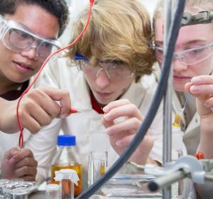
Biomedicine
The course
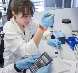
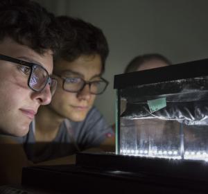
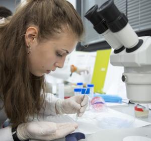
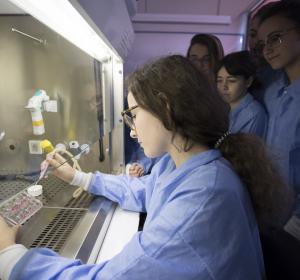
The Biomedicine course from the Crazy about Science programme of the Catalunya La Pedrera Foundation is a course developed by the Institute for Research in Biomedicine (IRB Barcelona) for A-Level students who want to explore some of the fascinating discoveries currently being made in the life sciences. Through this course, students will have the opportunity to deepen their knowledge of scientific theory and techniques in the field of biomedicine. They will work together with young researchers to experience how science is done at an international research institute, gain practical experience in the latest cutting-edge methodologies, and position themselves for a possible career in the life sciences.
The course will be held on different Saturdays between January and November 2024 at IRB Barcelona's facilities.
This course combines theory sessions and hands-on experimental activities, which will be held over 18 Saturdays a year. The course will cover 12 current scientific topics, from cellular and molecular biology to structural and computational biology and chemistry, presented by young researchers at IRB Barcelona. The course will be divided into two semesters (January-April and September-November) and each will cover 6 different topics. Each semester will have the same structure: during the first three Saturdays theoretical sessions will be held to examine each of the six research projects, and during the following six Saturdays the participants will be divided into groups of 4 people who will go into the laboratories for the practical sessions. So the groups will rotate and every Saturday a different practical session will be based on a real research project of IRB Barcelona and with the help of a young researcher who will tutor them for that particular experiment.
Participating students must commit to attending all course sessions throughout the year.
Specific objectives
Academic studies
- To foster among young people the scientific vocations linked to the world of biomedical sciences, helping to guide their academic and professional future.
- It provides the opportunity for your A-Level Research Project to be tutored by a leading research centre.
Research
- Bringing students closer to biomedical research.
- Understand the importance of basic research and its benefits to society.
- Understand the importance of multidisciplinarity.
- Discover the operation of the research centres linked to biomedicine.
Personal skills
- Working in teams.
- Promote critical thinking.
- Learn to present and communicate science in an understandable way.
Sessions 2024
1. From the lab bench to the microscope: observing the building blocks of our cells.
Session by: Nikolaos N. Giakoumakis (Advanced Digital Microscopy Facility)
Language: english.
Seeing is believing, and modern microscopy transformed the way that we can see the world around us. In our pursuit to comprehend complex dynamic cellular processes, it was necessary to surpass the physical limitations of our eyes. Thus, a few hundred years ago, we developed a scientific instrument to peer into the micro-cosmos around us. Light microscopy helped us discover the existence of microorganisms, shed light on the structure and the components of cells, and study the smallest parts of plants, fungi, and animals. In hospitals, microscopes help to diagnose by magnifying patients’ blood and tissue samples. Recent technological advances in computer sciences, physics and chemistry paved the way to re-revolutionising microscopes. Super-resolution microscopy is a rapidly developing field that consists of various fluorescence imaging techniques with the capacity to resolve objects below the classical diffraction limit of optical resolution. This field has given us the ability to observe the parts of subcellular structures in fixed or live samples in unprecedented detail.
In this course, students will be introduced to the principles of confocal microscopy and different super-resolution techniques. They will have the chance to prepare a sample suitable for fluorescence microscopy in order to observe the nuclei, the mitochondria and the cytoskeleton of cells in confocal and super-resolution microscopes.
2. Destroy to discover the cure – Investigating Angelman syndrome
Session by: Karen Sfyaki (Targeted Protein Degradation and Drug Discovery Laboratory)
Language: english.
Angelman syndrome is a complex genetic disorder affecting primarily the brain. The main characteristics of this syndrome are delayed development, speech impairment, and problems with movement and balance and, in severe cases, epileptic seizures, and sleep disorders. Currently, there is no cure for Angelman syndrome, but we know that it is caused by the loss of a protein called UBE3A. This protein belongs to the family of E3 ubiquitin ligases. These enzymes mark proteins for degradation through the proteasome. The proteasome is the cell’s trash machinery that breaks down the proteins that are damaged or that are no longer needed. Therefore, without UBE3A, its substrates cannot be degraded by the proteasome and it will accumulate. However, the substrates of UBE3A are not yet known and they could be potential therapeutic targets for Angelman syndrome.
In recent years, a new application of the proteasome system has been developed to study proteins. Scientists have strategically managed to use the proteasome to their advantage by tricking it into degrading proteins that they are interested in. This approach is known as “targeted protein degradation”. To achieve this, we add a degradation tag to the protein of interest. This tag can be recognised by drugs called degraders, which bring the target protein and an E3 ligase closely together, so that the protein can be marked for proteasomal degradation. This technology allows us to destroy any protein of interest and discover its downstream targets.
In this course, students will add the degradation tag to UBE3A using molecular cloning techniques and become familiar with DNA quantification, cloning and bacterial transformation.
3. The art of science across scales: from spins to cells
Session by: Borja Mateos (Molecular Biophysics Laboratory)
Language: english.
The term 'scientist' was first coined in 1834 by William Whewell and his friends in Cambridge. In analogy to the term 'artist', the new term aimed to replace previous designations that described the job of teaching and conducting research in scientific disciplines. In fact, it remains valid to this day that great science involves a great art. The ability to reflect curiosity and creativity in one's work is of utmost importance in our profession and has significant implications for various aspects of society. The scientific method provides the framework to channel curiosity into well-defined questions and employ creativity in the necessary methodology. To address fundamental questions, it is typically necessary to characterize our system of study at various temporal and spatial scales, ranging from microseconds to years and from Angstroms to meters. Accomplishing this requires the integration of multiple disciplines, both in vitro and in vivo, as well as the expertise of numerous scientists.
Biophysics encompasses a wide range of techniques aimed at addressing fundamental questions in biomedicine, drawing upon physics, chemistry, and biology. In this course, students will learn how to employ their questions and curiosity to formulate hypotheses and design experiments. Specifically, they will gain an understanding of how a diverse set of in vitro experiments can contribute to unraveling the molecular mechanisms of diseases. This will involve the combination of atomic-resolution techniques, such as NMR spectroscopy with microscopy, and various analytical assays. As an illustrative example, the course will explore the significant role played by intrinsically disordered proteins in regulating cellular fate and potentially triggering pathogenic processes.
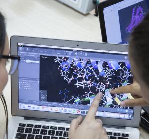
4. From biomedicine to computational biology
Sessió by: David Farré (Molecular Modelling and Bioinformatics Laboratory)
Language: english.
Interactions between small molecules are essential to living organisms. Amongst these molecules we find proteins and nucleic acids, whose interaction plays a crucial role in biology (e.g., in the storage, repair, expression and regulation of genetic information).
Protein-DNA interaction is defined mostly by two mechanisms, namely a direct or base read out, or an indirect or shape readout. The former describes the ability of proteins to distinguish different binding modes. The latter focuses on the importance of certain protein and DNA sequences to adopt different 3D structures that determine the binding sites. The relevance of these two mechanisms varies from protein to protein, but it is crucial to understand the role of a protein’s 3D structure.
In this course, students will address the 3D structure of different proteins from a computational perspective. The structure of a protein is believed to govern its function. Once the shape of a protein has been unravelled, its role within the essential biological mechanisms can be determined, thereby providing scientists with key information for the development of new methodologies that work with the shape of a given protein.
5. Chemistry in a Click!: obtaining complex molecules through easy reactions
Session by: Ines Maria Garcia (Research Unit on Asymmetric Synthesis).
Language: english.
Chemists have always wanted to harness the power of molecular assembly to build complex molecules and obtain the widest range of applications. The logic behind this interest is simple, as we are always in need of new molecular properties. These properties can be obtained from the joining of small molecular building blocks, which becomes more difficult as the complexity of the molecules increases.
In 2001, Barry Sharpless introduced the concept of Click Chemistry as a new approach to synthesise complex drug-like molecules by using a few practical and reliable reactions. Click Chemistry includes chemical reactions that are performed with high selectivity, high yield, and fast reaction rate, so that the “click” refers to the convenience afforded by snapping two objects together, like a luggage strap connection.
Click Chemistry has the following advantages over other types of reactions: it occurs in one pot; it is not disturbed by water; it generates minimal and usually harmless byproducts; and it has a high reaction specificity. Given these properties, this type of chemistry has been highly successful in recent years, leading its authors to won the Nobel Prize in Chemistry in 2022.
In this course, students will learn the basics of Click Chemistry and how to prepare these reactions. They will control these reactions using thin layer chromatography, detect the presence of both the reagents and the reaction products, and then finally purify the desired product.
1. Dealing with Stress – Learning survival mechanisms from the simplest eukaryotic organism
Session by: Maria Quintana & Guillem Pérez (Cell Signalling Laboratory)
Language: english.
A fundamental property of living cells is the ability to sense and respond to fluctuations in their environment. Cells have developed a large number of signal transduction pathways that serve to adapt to these changes. In our lab, we aim to unravel how cells detect, respond and adapt to environmental variations.
Working with the budding yeast Saccharomyces cerevisiae, a well-established eukaryotic model organism has several advantages: ease of manipulation, short life span, ability to produce a large number of offspring, and a sequenced genome. We use S. cerevisiae to study the adaptive eukaryotic responses to a variety of environmental stresses. Remarkably, many processes initially discovered and studied in yeast, such as transcription regulation, stress-signalling transduction and cell cycle regulation are conserved in higher eukaryotes.
Yeast and mammals share a conserved family of proteins known as Stress-Activated Protein Kinases (SAPKs) that sense and respond to environmental changes. The activation of SAPKs leads to a set of adaptive responses that involve the modulation of several physiological processes like changes in gene transcription, cell metabolism, protein translation, and cell cycle progression.
In this course, students will learn how to manipulate and work with S. cerevisiae to understand how cells detect and respond to external signals in order to ensure survival. For this purpose, we will study the activation of Hog1 SAPK upon different stress conditions and examine the cell fitness of various mutant strains when subjected to environmental insults.
2. Mirror, Mirror on the Wall… Who Is the Most Helpful Insect of Them All? – Getting to Know Tumorigenesis Through the Fly
Session by: Kaustuv Ghosh (Development and Growth Control Laboratory).
Language: english.
The fruit fly Drosophila melanogaster has been widely used as a model organism for more than one hundred years to address biological questions in various fields. This organism has emerged as a potent tool for genetic manipulation, offering innumerable possibilities to analyse the detailed interaction between cells and tissues.
One question could be raised: how can a tiny organism like D. melanogaster be used to understand the pathways of a disease as complex as cancer? The answer is quite simple. Many biological mechanisms are well conserved across evolution, including those related to pathological conditions. This conservation allows scientists to translate the knowledge acquired from less complex organisms to more complex ones. For instance, 75% of the genes related to human diseases and 68% of the genes related to human cancer have a counterpart in the fly. We are more similar to the fly than what we may think!
In this course, students will learn how to study the complex process of tumorigenesis using this model organism, both with a local and systemic approach. We will focus mainly on carcinomas, the most common type of tumour diagnosed in humans.
Carcinomas are derived from epithelial tissue, such as the skin, and they can become invasive or metastatic by spreading beyond the primary tissue layer and surrounding tissues or organs. In aggressive cancer cells, this transition is mediated by the activation of the EMT (Epithelial to Mesenchymal Transition) program, which causes the cells to undergo morphogenetic alterations that increase invasive capacity.
In this course, students will be introduced to the first steps in fly genetics. We will cross flies, identify genetic markers and balancer chromosomes, and apply other genetic tools that we use in the lab every day in order to generate tumorigenic flies and study them. Students will also learn about the anatomy of the fly in adult and larvae stages and use advanced microscopy to see several genetic markers in the tumoral tissues. Finally, students will perform in vivo dissections and their respective immunohistochemistry.
3. NMR to study molecules in solution
Sessions by: Miriam Condeminas (Structural Characterization of Macromolecular Assemblies).
Language: english.
Structural Biology aims at understanding the shape and function of biological molecules to decipher the basic mechanisms of life. It can also help identify molecules with pharmacological applications to treat human diseases. Nowadays, such questions can be answered using a wide variety of complementary tools that include Nuclear Magnetic Resonance (NMR).
In NMR, samples are irradiated with radio waves while placed inside a strong magnetic field. As a result, frequency changes in the nuclei of the molecules can be recorded and the corresponding signals plotted into NMR spectra. By combining various types of NMR experiments, each signal can be linked to a particular nucleus and thus yield important information regarding its chemical environment. Given that these experiments can be performed in aqueous solutions, NMR is very versatile in that it enables the observation of macromolecular flexible regions, as well as dynamic processes (invisible using other techniques), and it can also map binding interfaces.
In this course, students will learn the fundamentals of NMR, how to interpret the spectra resulting from various experiments, and the workflow required for assigning a peptide.
4. Using the Protein-Eating Machine to Treat Cancer
Session by: Jake Antony Ward (Targeted Protein Degradation and Drug Discovery Laboratory)
Language: english.
The control of the concentration, structure and location of proteins is important for the health and fitness of our cells. When proteins are damaged or are no longer needed, they are broken down within the cell in a process known as degradation. One of the main cellular systems involved in degrading unwanted or damaged proteins is called the proteasome. The proteasome is a large structure made up of many enzymes, such as proteases, which recognise and break down these proteins. For a damaged or unwanted protein to be recognised and broken down by the proteasome, a specific label, known as ubiquitin, is usually added to the protein. Certain enzymes known as ubiquitin ligases are involved in this tagging process.
In recent years, drugs have been developed that use this proteasome system in our cells to degrade a certain protein. This is an exciting new strategy, as it can allow us to the remove a given protein from a tumorogenic cell. This approach is known as Targeted Protein Degradation (TPD). This technology works by using a drug that brings the ubiquitin ligase and the target protein for degradation closer together. The protein is thus labelled with the ubiquitin-tag and is recognised by the proteasome and destroyed.
In this course, students will focus on a type of cancer known as Gastrointestinal Stromal Tumour (GIST). This cancer typically develops in the muscle cells of the gut. When mutated, the KIT protein is the molecule responsible for the formation and growth of this type of cancer. We are interested in designing drugs using the TPD strategy to remove KIT from GIST cancers. This line of research is exciting as it may lead to new treatments for GIST. Students will gain hands on experience of the key techniques of gel electrophoresis and Western blotting to monitor the degradation of KIT in human cancer cells.
5. Machine learning to fight drug resistance
Session by: Gema Rojas (Structural Bioinformatics and Network Biology Laboratory)
Language: english.
Drug resistance, which occurs when previously vulnerable cells become unresponsive to specific drugs, poses a significant public health concern worldwide. Antimicrobial resistance (AMR) exemplifies this issue, with estimates indicating that the annual death toll resulting from AMR could skyrocket from 700,000 to 10 million people by 2050. Similarly, drug resistance impacts the efficacy of cancer treatment, as patients may develop resistance following a relapse. The primary driver of drug resistance involves mutations in the DNA sequence of proteins associated with the drug's mode of action.
To comprehensively study this phenomenon, a crucial step involves analysing DNA sequencing data and establishing connections with protein structure and function to enhance our understanding of resistance mechanisms and devise strategies to overcome them. Machine learning has emerged as a valuable tool in predicting drug resistance by leveraging sequencing data.
In this course, students will have the opportunity to explore public databases and extract relevant biological information concerning drug resistance. They will work hands-on with sequencing data to understand the significance of mutations and observe how alterations in the sequence impact the shape and function of proteins. Finally, they will build a small machine learning model to predict drug resistance on the basis of sequence data.
Collaborating entities
The Institute for Research in Biomedicine (IRB Barcelona) is an internationally renowned research centre dedicated to understanding fundamental questions about human health and diseases.
Research at IRB Barcelona is carried out by 26 groups working in common with the common goal of carrying out multidisciplinary projects that address important biomedical problems affecting our society.
The goals of the institute include conducting multidisciplinary research of excellence in a framework between Biology, Chemistry and Medicine, offering its members, students and visitors high-level training in the field of the biosciences. We also provide an innovation component through active technology transfer as a benefit to society, as well as engaging in open dialogue with the public through engagement and education activities.
Other courses
Nutrition
Do you want to find out more about nutrition and the impact it has on our health?
Sustainability
Do you want to help to solve the challenges that the Earth will face in the next century?
Supercomputing
Would you like to work with the most powerful supercomputer in the country?
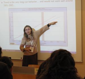
Economics
Do you want to analyse and understand some of the most important current economic phenomena?
Maths
Do you want to practice the intellectual challenge of abstract and creative mathematical thinking?
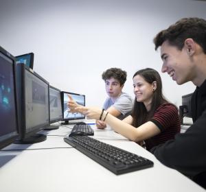
New Technologies
Are you interested in web development and programming, mobile applications, robotics or video games?
Chemistry
Are you passionate about chemistry and want to know first-hand how you work at an international research centre?
Physics
Would you like to get a closer look at the job of physics researchers?
Bioengineering
Are you are interested in the latest advances in bioengineering, biomaterials, nanobiotechnology and regenerative therapies?
Biochemistry
Would you like to learn what the latest biotechnological applications are that affect our lives?